
About Workshop
Spatial Biology: Immersive 3D Visualization of Cellular Structure is a cutting-edge workshop focused on 3D cellular imaging and spatial biology. Over three days, participants will explore advanced microscopy techniques, data integration, and AI-driven analysis to visualize and model complex cellular structures. Using industry-standard software like Imaris, Fiji, and 3D Slicer, attendees will gain hands-on experience to apply 3D imaging in drug discovery, disease research, and personalized treatments. This workshop is designed for professionals and researchers in biotechnology, bioinformatics, healthcare, and biomedical engineering seeking to enhance their expertise in advanced visualization and AI applications in cellular research.
Aim
The aim of this workshop is to provide participants with a comprehensive understanding of spatial biology and 3D visualization techniques, equipping them with hands-on skills to integrate AI with microscopy data for advanced analysis of cellular structures. The workshop will explore the latest tools and methods used in drug discovery, disease modeling, and personalized medicine, and offer practical applications in understanding the intricate biology of cells.
What Participants Will Learn
- To introduce participants to the core principles of spatial biology and its importance in understanding cellular structures and their roles in health and disease.
- To provide hands-on experience with 3D imaging techniques (Confocal, Electron, Super-Resolution, Light Sheet Microscopy) and tools used to analyze cellular structures.
- To demonstrate the integration of multi-omics data (RNA, protein, and genomic data) into 3D visual models for comprehensive biological analysis.
- To teach how to apply AI-driven machine learning models for feature extraction, classification, and anomaly detection in 3D cellular datasets.
- To explore the clinical applications of 3D cellular models in drug testing, cancer research, and personalized medicine.
Structure
Day 1: Introduction to Spatial Biology and 3D Visualization Techniques
- Introduction to spatial biology and its role in understanding cellular structures and functions.
- Key imaging techniques: Confocal, electron, super-resolution, and light sheet microscopy.
- Data integration: Combining multi-omics data (RNA, protein) with 3D imaging for comprehensive biological analysis.
- Applications of spatial biology in drug discovery, cancer research, and tissue engineering.
- Overview of tools and software for 3D visualization and image analysis.
Hands-On:
- Exploring 3D Cellular Structures (Fiji, Imaris)
- Preprocessing and Image Alignment (Imaris, 3D Slicer)
Tools Covered: Fiji, Imaris, 3D Slicer
Day 2: Advanced 3D Visualization and Data Interpretation
- Advanced microscopy techniques: Immunofluorescence, 3D microscopy, and fluorescent protein tags.
- How to integrate spatial transcriptomics and proteomics with 3D visualization.
- Mapping cellular pathways and understanding protein interactions using 3D models.
- The role of machine learning in automating the analysis of 3D cellular data.
- How 3D imaging contributes to personalized medicine and disease modeling.
Hands-On:
- 3D Imaging Analysis (Zen Software, ImageJ)
- 3D Pathway Mapping (Cytoscape, Imaris)
Tools Covered: Zen Software, ImageJ, Cytoscape, Imaris
Day 3: Data Analysis, Model Integration, and Clinical Applications
- Integrating 3D imaging data with computational models to simulate cellular dynamics and drug responses.
- The role of machine learning in extracting features from complex 3D biological data.
- Clinical applications of 3D cellular models in drug testing and personalized medicine.
- Using AI-driven models for real-time decision-making in drug discovery and disease treatment.
- Future trends in spatial biology: Digital twins, tissue engineering, and regenerative medicine.
Hands-On:
- Model Simulation Using 3D Data (MATLAB, Python)
- 3D Drug Testing and Clinical Visualization (Imaris, 3D Slicer)
Tools Covered: MATLAB, Python, Imaris, 3D Slicer
Important Dates
Registration Ends
04:30 PM IST
Workshop Dates
2026-05-27
05: 30PM
05: 30PM
What You Will Gain
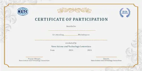
Outcomes
- Understand the fundamental concepts of spatial biology and how it contributes to drug discovery, disease research, and tissue engineering.
- Gain proficiency in using state-of-the-art 3D imaging tools like Imaris, Fiji, and 3D Slicer for cellular visualization and analysis.
- Learn to integrate multi-omics data (RNA, protein, genomic) with 3D imaging for deeper insights into cellular behavior and pathology.
- Be able to apply machine learning techniques to automate the analysis of complex 3D biological datasets.
- Be equipped to utilize 3D visualization for clinical applications such as drug testing, personalized medicine, and disease modeling.
Who Should Attend
- Academicians and researchers in biotechnology, bioinformatics, pharmaceuticals, biomedical engineering, and related fields.
- Industry professionals working in drug discovery, personalized medicine, healthcare, and biotech sectors.
- PhD researchers and postdoctoral fellows seeking to enhance their knowledge of 3D cellular imaging and AI applications in biological research.
- A basic understanding of cell biology, microscopy techniques, and data analysis is recommended but not required.
- Ideal for those seeking practical skills in 3D visualization, machine learning applications, and spatial biology for translational research.