
About Workshop
Explore AI-driven spatial biology using Vision Transformers, spatial transcriptomics, and multi-modal models for cancer microenvironment analysis.
Aim
To provide practical understanding of how Vision Transformers and multi-modal AI models can be used to analyze tissue images, spatial genomics data, and cancer microenvironment patterns for research and clinical translation.
Workshop Objectives
- Understand the role of AI in spatial biology and digital pathology.
- Learn how Whole-Slide Images are divided into patches for Transformer-based analysis.
- Generate attention maps to identify important tissue regions and immune cell patterns.
- Explore spatial transcriptomics and image-genomics data alignment.
- Apply Transformer models for gene marker prediction, tissue classification, and survival risk scoring.
- Create publication-ready visualizations such as heatmaps, UMAP plots, and Kaplan-Meier curves.
Structure
🗓️ Day 1: Preparing Tissue Topography for Transformers
- Understanding the shift from traditional pathology to AI-driven Spatial Biology.
- How Vision Transformers (ViTs) capture complex cell-to-cell environments where traditional CNNs fail.
- Step-by-step logic of breaking down Whole-Slide Images (WSIs) into patch sequences.
- Interpreting “Attention Maps” to identify hidden tumor-infiltrating immune cells.
💻 Hands-on Lab (Google Colab)
- Task: Convert a standard digital biopsy slide into spatial embeddings and extract feature sequences.
- Deliverable: Generate an interactive AI attention heatmap highlighting critical cellular regions.
🗓️ Day 2: Multi-Modal Fusion (Aligning Images with Genomics)
- Introduction to spatial transcriptomics data structures: mapping cellular morphology against gene expression profiles.
- Building Cross-Attention Transformer models to blend histology images with high-dimensional genetic data.
- Using Contrastive Learning frameworks to predict spatial gene expression directly from raw, low-cost H&E slides.
💻 Hands-on Lab (Google Colab)
- Task: Train a lightweight PyTorch Transformer layer to cross-reference tissue layouts with transcriptomic features.
- Deliverable: Map cross-modal tissue alignment and predict key cancer biomarkers from image inputs alone.
🗓️ Day 3: Clinical Translation & Cancer Microenvironment Profiling
- Mapping cellular “neighborhoods” and identifying immunosuppressive tumor boundaries.
- Utilizing Multiple Instance Learning (MIL) Transformers for whole-slide clinical classification.
- Translating AI-generated spatial biomarkers into publishable, peer-reviewed figures.
💻 Hands-on Lab (Google Colab)
- Task: Run a spatial transformer to process global WSI metrics for patient prognosis forecasting.
- Deliverable: Calculate patient survival risk scores and automatically generate a publication-quality Kaplan-Meier curve using Python.
Important Dates
Registration Ends
August 6, 2026 IST 4:30 PM
Workshop Dates
August 6, 2026 IST 4:30 PM
What You Will Gain
- Live & recorded sessions
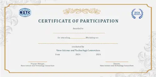
- e-Certificate upon completion
- Post-workshop query support
- Hands-on learning experience
Who Should Attend
- Students in biotechnology, bioinformatics, biomedical science, and related fields
- PhD scholars and researchers working in cancer biology, pathology, computational biology, or spatial biology
- Academicians and faculty members interested in AI-driven digital pathology and precision medicine
- Professionals from healthcare AI, biomedical research, diagnostics, and precision oncology domains
- Learners interested in Vision Transformers, spatial transcriptomics, and cancer microenvironment analysis