
About Workshop
This three-day international workshop provides practical training in AI-based radiology using CT, MRI and X-ray data. Participants will learn medical image preprocessing, multimodal data fusion, MedSAM segmentation, Vision Transformer models, chest X-ray classification and clinical explainable AI.
Hands-on Google Colab sessions will use MONAI, MedSAM, Swin UNETR, TorchXRayVision, PyTorch and Captum to build reproducible medical imaging workflows.
Aim
To develop practical skills in building, evaluating and explaining AI models for multimodal radiology and medical image analysis
What Participants Will Learn
Participants will learn to:
- Process DICOM and NIfTI medical images.
- Normalize, align and resample CT and MRI scans.
- Build multimodal medical imaging pipelines.
- Apply MedSAM and Swin UNETR for segmentation.
- Perform multi-label chest X-ray classification.
- Generate Grad-CAM and Integrated Gradients explanations.
- Evaluate models using Dice Score, AUROC, precision, recall and F1-score.
- Prepare publication-ready figures and validation reports.
Structure
🗓️ Day 1: Multi-Modal Radiology Data Pipeline Engineering
- Objective: Build standardized and memory-efficient pipelines for processing multi-modal medical imaging data.
- Introduction to CT, MRI, X-ray, and clinical data fusion.
- Understanding DICOM and NIfTI medical imaging formats.
- Managing LPS/RAS orientation and spatial alignment.
- Applying intensity normalization and Hounsfield Unit windowing.
- Comparing early-fusion and late-fusion architectures.
- Isotropic voxel resampling for consistent 3D analysis.
- Patch sampling and sliding-window inference for large volumes.
🛠️ Hands-on Lab (Google Colab)
- Task: Build a MONAI pipeline to load, normalize, resample, and spatially align paired T1/T2 MRI and CT scans.
- Tools Covered: MONAI, SimpleITK, PyTorch, TorchIO, and Google Colab.
🗓️ Day 2: Vision Transformers, MedSAM and Automated Pathology Detection
- Objective: Apply transformer architectures and medical foundation models for segmentation and multi-label diagnosis.
- Evolution from U-Net to transformer-based medical image segmentation.
- Architecture and clinical applications of Swin UNETR.
- Introduction to MedSAM and medical foundation models.
- Using bounding-box, point, and mask-based segmentation prompts.
- Zero-shot segmentation of lesions and anatomical regions.
- Transfer learning with pre-trained radiology models.
- Multi-label detection of pneumonia, pneumothorax, and pulmonary nodules.
🛠️ Hands-on Lab (Google Colab)
- Task: Segment lung nodules using MedSAM prompts and perform multi-label chest X-ray classification using TorchXRayVision.
- Tools Covered: MedSAM, TorchXRayVision, Swin UNETR, MONAI, Hugging Face, and Google Colab.
🗓️ Day 3: Explainable AI, Clinical Validation and Research Publication
- Objective: Interpret medical AI predictions, evaluate model performance, and prepare reproducible research outputs.
- Understanding the importance of explainability in clinical AI systems.
- Creating Grad-CAM and Layer-CAM visualizations.
- Applying Integrated Gradients to multi-modal medical imaging models.
- Evaluating segmentation using Dice Score and Hausdorff Distance.
- Evaluating classification using AUROC, precision, recall, and F1-score.
- Designing ablation studies and comparative experiments.
- Creating publication-ready figures and reproducible research workflows.
🛠️ Hands-on Lab (Google Colab)
- Task: Generate saliency maps using Captum and Grad-CAM, create 300-DPI radiology visualizations, and produce statistical validation reports.
- Tools Covered: Captum, PyTorch Grad-CAM, scikit-learn, MONAI Label, Matplotlib, Seaborn, and Google Colab.
Important Dates
Registration Ends
4:30 PM
Workshop Dates
2026-08-20
5:30 PM
5:30 PM
What You Will Gain
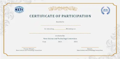
Outcomes
By the end of the workshop, participants will be able to:
- Build standardized MONAI imaging pipelines.
- Segment lesions and anatomical regions using MedSAM.
- Apply Vision Transformers to medical imaging tasks.
- Classify radiological abnormalities from chest X-rays.
- Interpret model predictions using clinical XAI methods.
- Evaluate segmentation and classification performance.
- Create reproducible research workflows and publication-ready outputs.
Who Should Attend
- Radiologists and clinical researchers
- PhD scholars and postgraduate students
- Academicians and faculty members
- Biomedical and medical imaging researchers
- Healthcare data scientists
- AI and machine learning professionals
- Biomedical engineers
- Medical imaging and diagnostic-AI professionals