
About Workshop
Aim
To develop practical skills in building, evaluating and explaining AI models for multimodal radiology and medical image analysis.
Participants will learn to:
- Process DICOM and NIfTI medical images.
- Normalize, align and resample CT and MRI scans.
- Build multimodal medical imaging pipelines.
- Apply MedSAM and Swin UNETR for segmentation.
- Perform multi-label chest X-ray classification.
- Generate Grad-CAM and Integrated Gradients explanations.
- Evaluate models using Dice Score, AUROC, precision, recall and F1-score.
- Prepare publication-ready figures and validation reports.
Structure
🗓️ Day 1: Multi-Modal Radiology Data Pipeline Engineering
Objective: Build standardized and memory-efficient pipelines for processing multi-modal medical imaging data.
- Introduction to CT, MRI, X-ray, and clinical data fusion.
- Understanding DICOM and NIfTI medical imaging formats.
- Managing LPS/RAS orientation and spatial alignment.
- Applying intensity normalization and Hounsfield Unit windowing.
- Comparing early-fusion and late-fusion architectures.
- Isotropic voxel resampling for consistent 3D analysis.
- Patch sampling and sliding-window inference for large volumes.
🛠️ Hands-On: Build a MONAI pipeline to load, normalize, resample, and spatially align paired T1/T2 MRI and CT scans.
🧰 Tools Covered: MONAI, SimpleITK, PyTorch, TorchIO, and Google Colab.
🗓️ Day 2: Vision Transformers, MedSAM and Automated Pathology Detection
Objective: Apply transformer architectures and medical foundation models for segmentation and multi-label diagnosis.
- Evolution from U-Net to transformer-based medical image segmentation.
- Architecture and clinical applications of Swin UNETR.
- Introduction to MedSAM and medical foundation models.
- Using bounding-box, point, and mask-based segmentation prompts.
- Zero-shot segmentation of lesions and anatomical regions.
- Transfer learning with pre-trained radiology models.
- Multi-label detection of pneumonia, pneumothorax, and pulmonary nodules.
🛠️ Hands-On: Segment lung nodules using MedSAM prompts and perform multi-label chest X-ray classification using TorchXRayVision.
🧰 Tools Covered: MedSAM, TorchXRayVision, Swin UNETR, MONAI, Hugging Face, and Google Colab.
🗓️ Day 3: Explainable AI, Clinical Validation and Research Publication
Objective: Interpret medical AI predictions, evaluate model performance, and prepare reproducible research outputs.
- Understanding the importance of explainability in clinical AI systems.
- Creating Grad-CAM and Layer-CAM visualizations.
- Applying Integrated Gradients to multi-modal medical imaging models.
- Evaluating segmentation using Dice Score and Hausdorff Distance.
- Evaluating classification using AUROC, precision, recall, and F1-score.
- Designing ablation studies and comparative experiments.
- Creating publication-ready figures and reproducible research workflows.
🛠️ Hands-On: Generate saliency maps using Captum and Grad-CAM, create 300-DPI radiology visualizations, and produce statistical validation reports.
🧰 Tools Covered: Captum, PyTorch Grad-CAM, scikit-learn, MONAI Label, Matplotlib, Seaborn, and Google Colab.
Important Dates
Registration Ends
Workshop Dates
What You Will Gain
- Live & recorded sessions
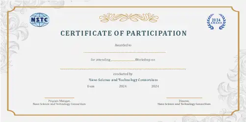
- e-Certificate upon completion
- Post-workshop query support
- Hands-on learning experience
Who Should Attend
- Radiologists and clinical researchers
- PhD scholars and postgraduate students
- Academicians and faculty members
- Biomedical and medical imaging researchers
- Healthcare data scientists
- AI and machine learning professionals
- Biomedical engineers
- Medical imaging and diagnostic-AI professionals