
About Workshop
Quantitative pathology and biomarker analysis play a critical role in precision medicine, enabling objective assessment of tissue and cellular features to inform disease diagnosis and treatment. Traditional manual analysis is often time-consuming and subject to variability. AI techniques, including machine learning and deep learning, provide scalable, accurate, and reproducible methods for quantitative analysis of histopathology images. This workshop will cover AI-driven approaches for automated tissue segmentation, feature extraction, and biomarker quantification. Participants will also explore how AI models can correlate image-derived features with clinical and molecular data to discover and validate novel biomarkers. Real-world case studies will demonstrate the impact of AI in improving disease diagnosis and personalized treatment planning.
Aim
This workshop aims to introduce participants to the application of Artificial Intelligence (AI) in quantitative pathology and biomarker analysis. Participants will learn how AI can automate image analysis, quantify tissue and cellular features, and identify disease biomarkers to improve diagnostics, prognostics, and personalized medicine strategies.
What Participants Will Learn
- Understand the principles of quantitative pathology and biomarker analysis.
- Learn how AI can automate tissue segmentation and feature quantification.
- Gain hands-on experience with deep learning models for image analysis.
- Explore AI-driven approaches for biomarker discovery and validation.
- Apply AI techniques to integrate pathology and clinical data for personalized medicine.
Structure
Day 1: Biomarkers & Image Feature
- Assays & scoring: IHC/ISH/mIF; H-score, Allred, CPS/TPS.
- WSI pipeline: scanning, tiling, TMAs vs WSIs, formats; data governance & PHI.
- Stains & QC: color normalization (Macenko/Reinhard), deconvolution; artifact/tissue detection.
- Segmentation & features: nuclei/cell/region; thresholding/watershed/StarDist/Hover-Net; morphology, intensity (DAB OD), texture (GLCM), spatial (Ripley’s K).
- Hands-on: QuPath navigation/ROI, deconvolution; CellProfiler nuclei features → export.
- Mini-task: compute H-score on patches across cases.
- Data & prep: patient-level splits, leakage control, stratification, imbalance fixes; tiling/augmentation, stain-aware transforms.
- Models: tabular ML (LR/RF/XGB), CNNs on tiles, MIL/attention for WSIs.
- Labels & scoring: pathologist/proxy/consensus, active learning; automated H-score/Allred, PD-L1 CPS/TPS; tumor vs stroma.
- Eval & robustness: patient-level metrics, calibration, error analysis; cross-site/stain variability, domain adaptation; tracking/audit.
- Hands-on: baseline on features, fine-tune CNN, simple MIL; per-case scores.
- Mini-task: compare classical vs deep models + calibration.
- Stats: group tests, covariate correlations, survival (KM/Cox); ROC/PR, thresholds, decision curves.
- Visualization: heat/attention maps, Grad-CAM, spatial positivity maps.
- Reporting & clinical: notebooks/figures/tables; pathologist-in-loop, LIS/PACS fit, CAP/CLIA.
- Deployment: packaging, batch vs real-time, sizing, monitoring, bias/fairness & drift; SOPs, validation, change control.
- Hands-on: cohort summaries, survival curves, explainability overlays; 1-page validation report.
- Mini-task: clinician-readable score report with interpretation.
Important Dates
Registration Ends
7:00 PM
Workshop Dates
2025-10-14
8:00 PM
8:00 PM
What You Will Gain
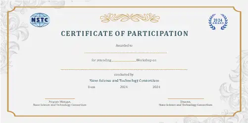
Outcomes
- Ability to apply AI for quantitative analysis of histopathology images.
- Skills in automating tissue segmentation and cellular feature extraction.
- Hands-on experience in AI-based biomarker discovery and validation.
- Understanding of integrating image-derived features with clinical and molecular data.
- Knowledge of AI applications in precision medicine and oncology.
- Enhanced ability to interpret AI outputs for clinical decision-making.
Who Should Attend
- Undergraduate/Postgraduate Degree in Bioinformatics, Biotechnology, Computational Biology, Biomedical Engineering, or related fields.
- Professionals in pathology, clinical research, oncology, or medical imaging.
- Data Scientists and AI/ML Engineers interested in applying AI to pathology and biomarker analysis.
- Individuals with a strong interest in AI applications in healthcare and precision medicine.